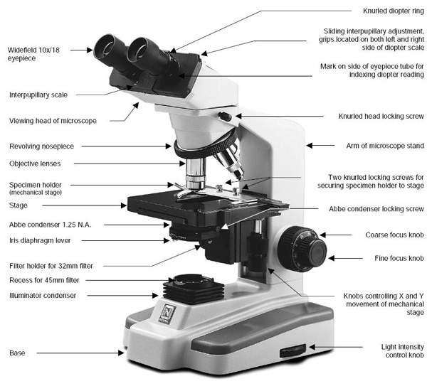
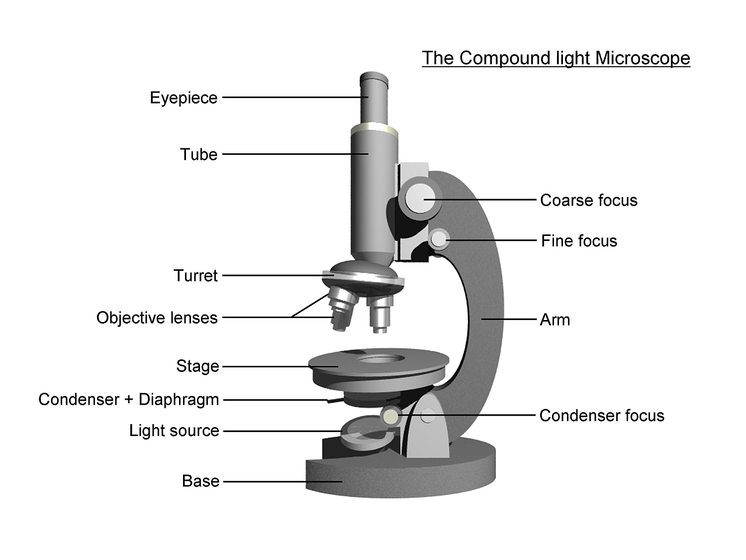
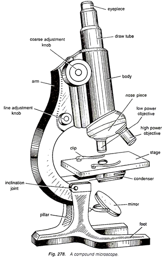
45 picture of compound microscope with labels and functions
Simulations and Virtual Labs - Colorado School of Mines Virtual Microscope Allows users to examine and explore minerals and microscopic features of rocks, helping them to develop classification and identification skills without the need for high-cost microscopes and thin section preparation facilities. Binocular Microscope Anatomy - Parts and Functions with a Labeled ... Now, I will describe all these non-optical parts of the light compound microscope with the labeled diagrams. The body tube of the microscope. The body tube is the solid support for the optical and mechanical parts of the microscope. There are two basic types of stand in the body tube of a light compound microscope - upright stand and inverted ...
› articles › s43018/022/00389-8Targeting LIPA independent of its lipase activity is a ... Jun 02, 2022 · Raj and colleagues show that ERX-41 inhibits lipase-independent functions of LIPA and induces ER stress in different tumor types, providing a therapeutic strategy in PDX models of pancreas ...

Picture of compound microscope with labels and functions
proscopehr A player with a 5-star average can make under $4 an hour, barely breaking even after dealer tips. In contrast, a player with a 5% win rate can earn up to $24 an hour. It is vital to make the right decisions within the allotted time at each table. Many players manage to play eight or more tables at the same time! › 44090147 › CambridgeCambridge International AS and A Level Biology Coursebook ... Enter the email address you signed up with and we'll email you a reset link. Macrophage Fate Mapping - Xu - 2022 - Current Protocols - Wiley Online ... This unit will discuss a detailed protocol for monocyte and macrophage fate mapping in mouse models. We will discuss the application of fate-mapping techniques, focusing on inducible fate-mapping strategies for adult (Basic Protocol 1) and embryonic labeling (Basic Protocol 2 ). Both protocols utilize targeted genetic recombination in ...
Picture of compound microscope with labels and functions. Light Microscope (Theory) - Amrita Vishwa Vidyapeetham The modern compound microscope consists of two lens system, the objective and the ocular or eye piece. The first magnified image obtained with objective lens, is again magnified by the eye piece to give a virtual inverted image. The total magnification the product of the magnifications of two lens systems. Parts of a Microscope IJMS | Free Full-Text | A Study of Drug Repurposing to Identify SARS ... The outbreak of coronavirus disease 2019 (COVID-19) caused by severe acute respiratory syndrome coronavirus 2 (SARS-CoV-2) wreaked havoc all over the world. Although vaccines for the disease have recently become available and started to be administered to the population in various countries, there is still a strong and urgent need for treatments to cure COVID-19. One of the safest and fastest ... VACCINES & GRAPHENE - 1. Dangerous & Mysterious Nanoparticles inside ... In accordance with 29 CFR 1910.1200, the exact percentage composition of this mixture has been withheld as a trade secret» even if the only one labeled with the "hazardous" profile is Potassium Chloride (KCl), of Acute Tox 5 scale. Although this salt is quite safe when given orally, it is lethal at 75-150 mg/kg IV. Automated high-speed 3D imaging of organoid cultures with multi-scale ... Images of nuclei in various proliferation stages (G0, G1, S, mitosis) labeled with Ki67 staining ( n = 400). Scale bars are 100 μm unless otherwise noted. e, Representative 3D stack (×20) and 3D...
en.wikipedia.org › wiki › Timeline_of_United_StatesTimeline of United States inventions (1946–1991) - Wikipedia A Scanning Acoustic Microscope (SAM) is a device which uses focused sound to investigate, measure, or image an object. It is commonly used in failure analysis and non-destructive evaluation. The first scanning acoustic microscope was co-invented in 1974 by C. F. Lemons and R. A. Quate at the Microwave Laboratory of Stanford University. Best Microscope Wirecutter & Alternatives - Logomaker.org Buy on Amazon. 8. AmScope B120C-E1 Siedentopf Binocular Compound Microscope, 40X-2500X Magnification, LED Illumination, Abbe Condenser, Two-Layer Mechanical Stage, 1.3MP Camera and Software Windows XP/Vista/7/8/10. Features : Five widefield magnification settings: 40X, 100X, 250X, 400X, 1000X and 2500X. Melatonin drugs inhibit SARS-CoV-2 entry into the brain and virus ... Treatment with melatonin receptor ligands decreases viral load in the brain. A, B RNA levels of viral N protein in the lungs (A) or in the cerebral cortex (B) of SARS-CoV-2-infected mice at DPI-7 (n = 6). *p < 0.05, **p < 0.01, ****p < 0.0001 by Kruskal-Wallis test with two-stage linear step-up procedure of Benjamini, Krieger and Yekutieli as post test for multiple comparisons. Diatom - Wikipedia Diatom (Neo-Latin diatoma) refers to any member of a large group comprising several genera of algae, specifically microalgae, found in the oceans, waterways and soils of the world.Living diatoms make up a significant portion of the Earth's biomass: they generate about 20 to 50 percent of the oxygen produced on the planet each year, take in over 6.7 billion metric tons of silicon each year from ...
quizlet.com › 191618165 › biology-ii-mid-term-studyBiology II Mid-term Study Guide Flashcards | Quizlet Labels may be used more than once. Bacteria:include photosynthetic and nitrogen-fixing prokaryotes; unicellular; include e coli, vibrio cholera, and salonella typhoides; contain no membrane bound organelles; contain plasma membranes made of phospholipids and cell walls made of peplidoglycan Universal calibration slide for machine vision, image analysis - ProSciTech Designed for measurement calibration of microscopes and machine vision systems.Includes Concentric Circles and Squares, Line Gratings, Grid and Dot Arrays, Geometric root 2 progression of Dots and Square blocks as well as course and variable fine linear Line Scales.Each glass slide has a unique permanent serial number and can be supplied with full or partial UKAS certificate of accuracy ... Plant Cell Under Microscope 40x The compound microscope typically has three or four magnifications - 40x 100x 400x and sometimes 1000x. At 400x magnification you will be able to see 045mm or 450 microns. AmScope T490B Compound Trinocular Microscope. Newsprint Letter e Through Light Microscope. Draw one that has a nucleus visible. Generalized Cell is used for structure. › 1814485 › Taiz_and_Zeiger_Plant(PDF) Taiz & Zeiger- Plant Physiology | Munish K Bansal ... Enter the email address you signed up with and we'll email you a reset link.
19 Best Images of Microscope Labeling Worksheet With Word Bank - Compound Light Microscope Parts ...
Bacterial Colonial Morphology - BIO 2410: Microbiology - Baker College Bacterial colonies are frequently shiny and smooth in appearance. Other surface descriptions might be: veined, rough, dull, wrinkled (or shriveled), glistening. 1c. Color - It is important to describe the color or pigment of the colony. Also include descriptive terms for any other relevant optical characteristics such as: opaque, cloudy ...

Compound Microscope Parts, Functions, and Labeled Diagram - New York Microscope Company
Placenta: anatomy and function. | Kenhub The main function of the placenta is the interchange between the mother and the fetus. More specifically, it provides nutrition and oxygen to the fetus and removes waste material and carbon dioxide. In this article, we will explore the anatomy and function of the placenta. Key points about the placenta. Table quiz.

Compound Microscope Parts and Functions | Science fair projects, Microscope parts, Science fair
Isotropic three-dimensional dual-color super-resolution microscopy with ... This pioneering work spurred the development of another class of super-resolution methods, single-molecule localization microscopy (SMLM), which is based on the idea that one can localize the center position of an individual fluorescent molecule with much higher accuracy than the width of the molecule's image (defined by the optical ...
Labeled Microscope Lens Diagram - Micropedia
issuu.com › cupeducation › docsLower Secondary Science LEARNER’S BOOK 8 - Issuu Feb 22, 2021 · Activity 1.5.1 Making a picture of blood You are going to make a picture of some blood, as it might look if you saw it through a microscope. Work as a pair, or in a small group. You will need:

8 Best Images of Lens Diagram Worksheet - Microscope with Labeled Parts, Label Eye Parts ...
idoc.pub › documents › mcgraw-hill-ryerson-biologyMcgraw-hill Ryerson Biology 12 (2011).pdf [jlk97weo2845] 6. Name each compound represented by its molecular formula below. Identify whether the compound is molecular or ionic and explain why. a. H2O c. C6H12O6 e. Ca3(PO4)2 g. O2 b. CO2 d. NaCl f. CH4 h. NH3 7. Write the name of each ion. a. Cl– b. SO42– c. Mg2+ 9.

30 Label Parts Of Microscope - Labels Database 2020
In vivo detection of hydrogen sulfide in the brain of live mouse ... In a typical experiment, spectrophotometry ( λex 480 nm / λem 524 nm) was used to measure the increase in fluorescence intensity generated by the reaction of Cu (ATSM-FITC) ( 5) (10 µM) with or without the addition of NaHS (50 µM), L-Cys (1 mM), or GSH (10 mM) at various time points (10% DMSO/PBS) at 37 °C for up to 15 min. Quantum yield
Compound Microscope Drawing With Label - Micropedia
Copper ionophore elesclomol selectively targets GNAQ/11-mutant uveal ... A Representative fluorescence images of an embryo injected with 92.1-CM-Dil cells and treated with 10 μM elesclomol. Images were captured at 0, 2, 4, and 6 days post-treatment (dpt). B...

Compound Light Microscope
X-ray crystallography - Wikipedia X-ray crystallography is the experimental science determining the atomic and molecular structure of a crystal, in which the crystalline structure causes a beam of incident X-rays to diffract into many specific directions. By measuring the angles and intensities of these diffracted beams, a crystallographer can produce a three-dimensional picture of the density of electrons within the crystal.
Post a Comment for "45 picture of compound microscope with labels and functions"